Morphological Feature Analysis of Retinal Pigment Epithelial Cells from Mice during Aging
| Advisor | Dr. Yi Jiang (Georgia State University) |
| Presented At | • Georgia State Undergraduate Research Conference (GSURC) 2025 (Poster) |
| Code Repository | GitHub: vanthienphan2004/RPE-Morphological-Analysis |
Abstract
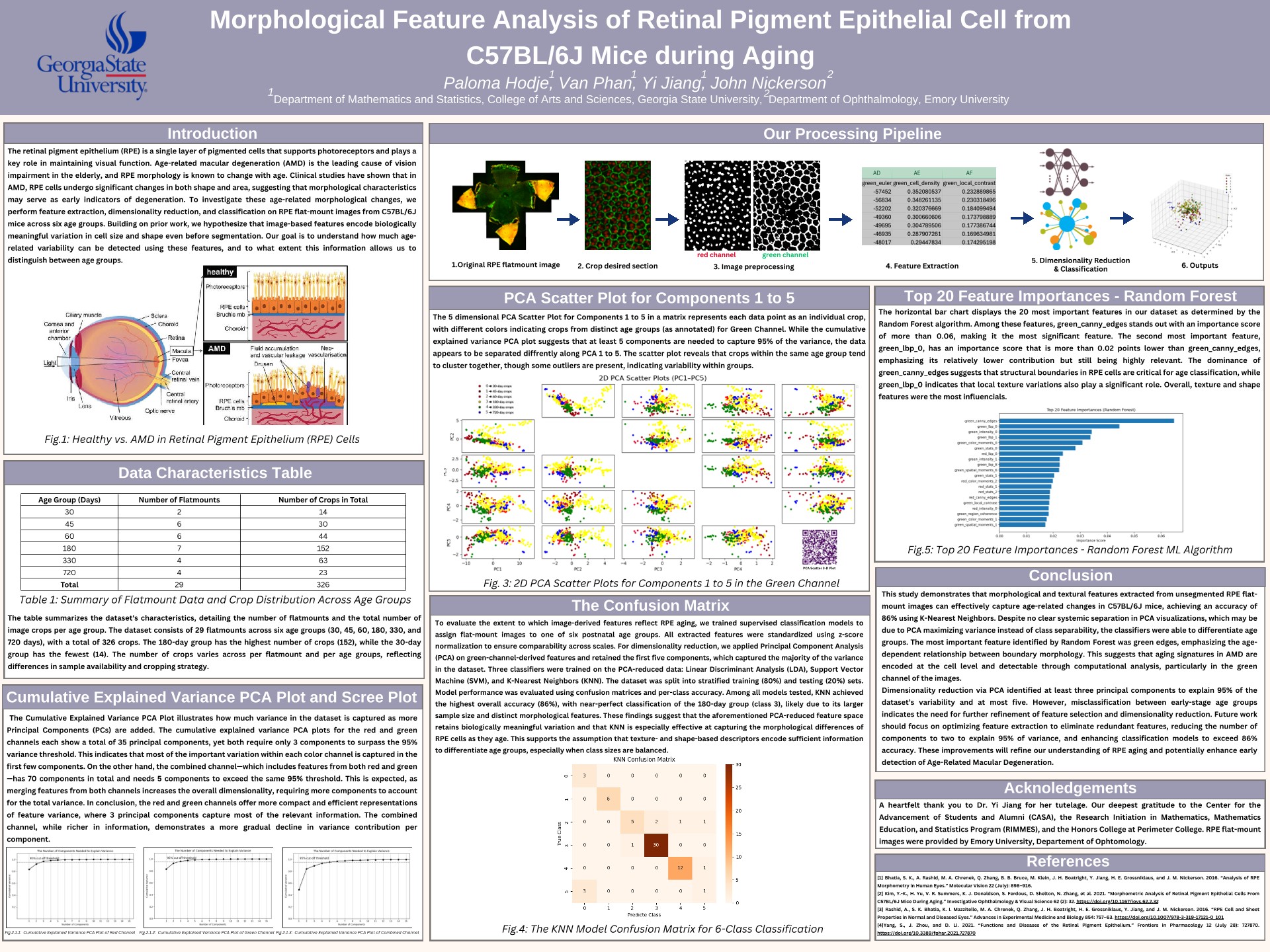
Age-related phenotypic degeneration of the Retinal Pigment Epithelium (RPE) plays a central pathological role in the progression of Age-related Macular Degeneration (AMD). Detecting subtle cellular morphological drift early in the aging process is critical for establishing preventive and therapeutic benchmarks.
In this work, we developed an automated, end-to-end computer vision and machine learning pipeline to systematically extract and evaluate high-dimensional cellular features from microscopic images of C57BL/6J mouse RPE cells. Using OpenCV and scikit-image, we computed 133 distinct morphological, geometric, and texture features across a dataset of 326 segmented RPE cell images.
A stacking ensemble architecture combining XGBoost, LightGBM, and CatBoost with a Logistic Regression meta-classifier achieved a 90%+ cross-validation F1-score in distinguishing aged from young cellular states. Feature importance analysis revealed that Local Binary Patterns (LBP) texture descriptors alongside geometric shape metrics (circularity and aspect ratio) served as the primary discriminating biomarkers, highlighting structural irregularity as a hallmark of early retinal epithelial aging.
Conference Poster